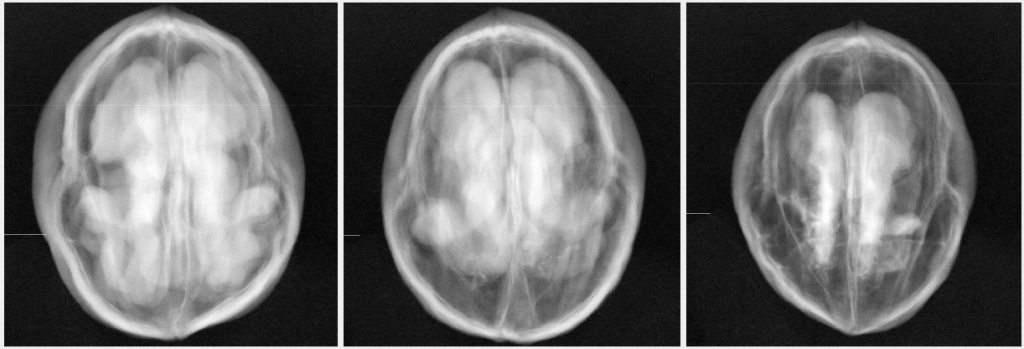
Nothing beats x-rays to find out what’s hidden inside opaque and dense objects. So, once we heard about the kernel shrivel problem we set out to try x-rays at once. Here’s a simplified report of our preliminary work, which we decided to put out there to look for potential interest in the industry.
X-ray imaging of walnuts in shell
We imaged 166 walnut samples, kindly provided by Quality Walnut Producers Pty Ltd. The imaging work has been done in the x-ray lab of Dr Marcus Kitchen at Monash University. We used a medical (mammography) x-ray unit and a flat panel digital detector. The x-ray unit was set to 35 kVp and 50 mAs. Walnut samples were placed in close proximity of the flat panel detector to acquire images. All walnut samples were placed in approximately the same orientation with respect to the detector.
Some result
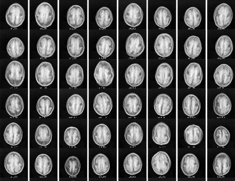
Here’s the image overview of some of the samples we measured. We made sure to have a wide range of size and shell colour. Moreover we used walnuts from different growing regions around Victoria and New South Wales.


